
There’s Something Fishy About that X-Ray
Countless animals arrive at the Zoo every year, each with their own story and background. Some arrive with long medical histories. But often, animals come to the Zoo with very little known about their medical backgrounds at all.
To help ensure each new animal receives the care and attention they need, all new Zoo residents receive a thorough intake exam.
These examinations are critical to the Zoo’s approach to preventive health care and can identify medical concerns before an animal even appears sick.
In August, a white-spotted puffer fish was donated to the Zoo by a private party. During the fish’s intake exam, standard radiographs were taken. The pictures revealed several findings that were not visible on external examination.
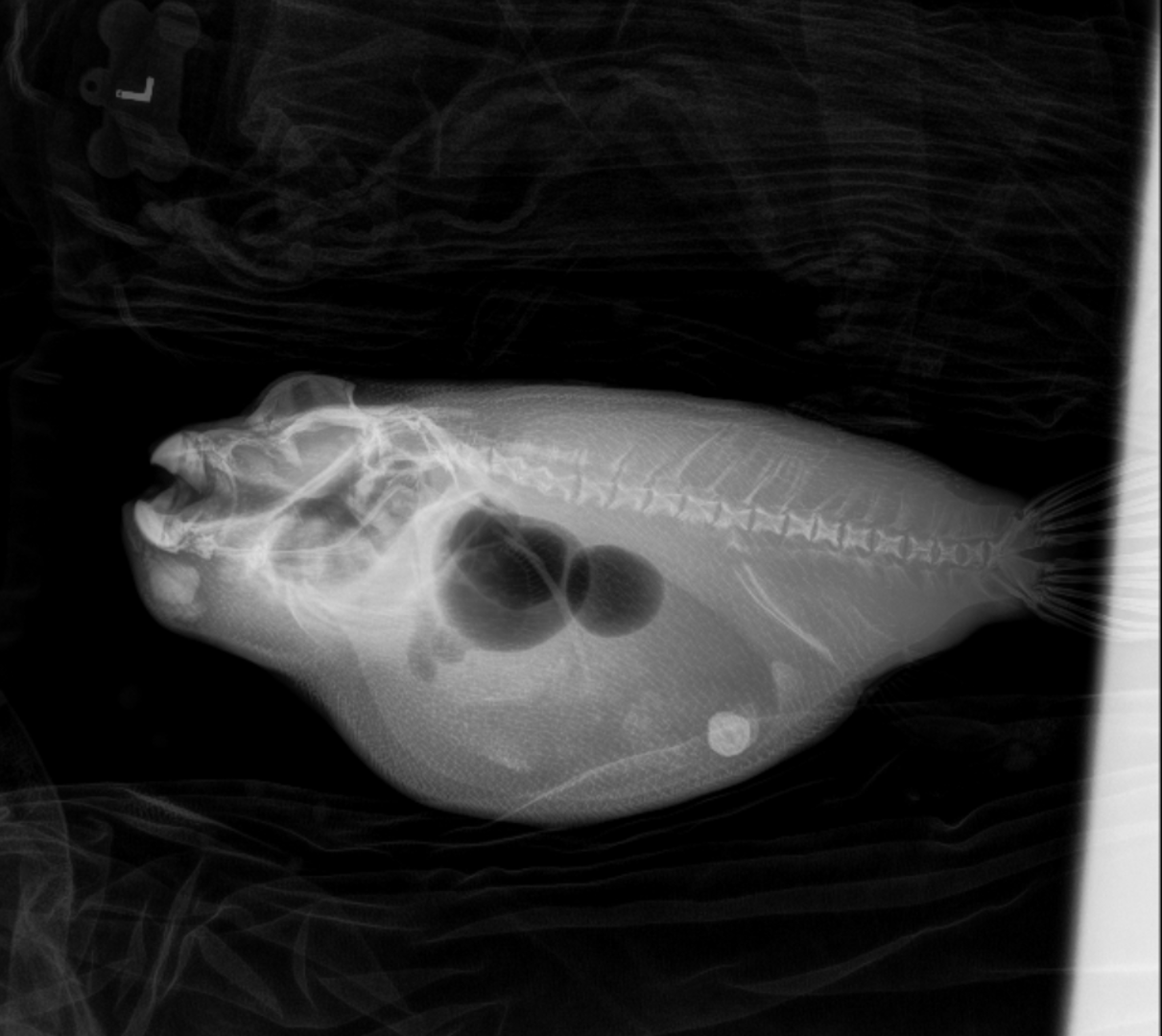
This white-spotted puffer fish’s radiograph reveals a stone in the stomach and a mass seen below the jaw.
A large stone was found in the puffer’s stomach, as well as a mass-like structure beneath the jaw. Following the x-ray imaging, an ultrasound was performed to collect more information. The fish is now scheduled for a follow-up procedure to remove the stone and biopsy the jaw mass to decide the best course of treatment.
This story is a clear example of why preventive care is so important, and why diagnostic imaging like radiographs are part of most intake exams. Without these regular exams, many animals that appear healthy may not receive the treatment they need for hidden conditions.
Proactive care like this is made possible by donors like you, who power wildlife-saving efforts for animals at the Zoo and their counterparts in the wild.